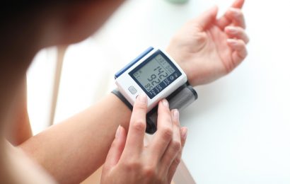
If you are lucky enough to have good eyesight, you may be surprised when your diabetes care team recommends that you make an appointment with an eye doctor. If your vision is stable, and your eyes don’t bother you, why should you have your eyes checked?
The answer is that many potentially devastating eye problems develop without causing discomfort or distorting vision. Glaucoma and cataract are examples of eye problems that occur commonly in older adults and more frequently in people with diabetes. Generally, these conditions are treatable, but if not caught early enough, they can lead to vision loss or even blindness.
In addition, there’s diabetic retinopathy, a serious complication that is more likely to occur in people with Type 1 diabetes but may develop in anyone with diabetes. Tight blood glucose control can significantly reduce the incidence and severity of diabetic retinopathy, but the only way to identify this and other eye problems in their earliest and most treatable stages is to have regular, comprehensive eye examinations.
— Keep an eye on your vision! Learn about preventive steps and treatments for diabetic retinopathy from retinal specialist Dr. Charles Wykoff. >>
There’s no reason to avoid an eye exam; it involves a series of painless tests that check your visual acuity and general eye health and screen for signs of disease. Before we discuss what to expect at the exam, let’s take a look at the eye and how it works.
The eye
The eye is a hollow organ about the size of a Ping-Pong ball, with an opening at the front that lets in light, and a gelatinous substance called vitreous filling most of the inside. It functions in a manner similar to a camera.
The aperture through which light enters the eye is the pupil, the black-seeming hole in the middle of the eye. The iris, the colored ring of muscle tissue surrounding the pupil, controls the amount of light coming in by narrowing or dilating the pupil. The “white” of the eye, or sclera, is a hard shield of tissue that encircles and protects the opening of the eye. A thin layer of tissue called the conjunctiva protects the sclera and connects the eye to the eyelid.
The eye’s main focusing element is the cornea, a clear, hard tissue covering the iris and the pupil. The curve of the cornea bends, or refracts, light rays, focusing them on the retina at the back of the eye. A pool of fluid called aqueous humor fills a cavity between the cornea and the iris. Directly behind the iris is the lens, an elastic disc about the size and shape of an M&M candy, which flexes to fine-tune focus.
Lining the back of the eyeball is the retina, a complex, photosensitive membrane of many layers. This is the “film” of the eye and its most important part. When light is focused onto the retina, photosensitive cells translate the light into electrical impulses, which are then sent via the optic nerve to the brain, where an image is formed. (See “Anatomy of the Eye” for illustrations.)
The eye exam
Like most doctor visits, the eye exam begins with a little paperwork. You will be asked to answer questions or fill out a form, providing information about your general health, any medicines you take, allergies or eye problems you have, and your family medical history. While some of this information may seem irrelevant, asking these routine questions is the only way to establish background information that really does matter. Having high blood glucose or even taking a common, over-the-counter medicine can cause fluctuations in your vision that might make a difference in your exam.
Background complete, the next step in most eye exams involves assessing your visual acuity, or how well you can see. Vision is measured by the size of the letters you can easily read on the eye chart, which is usually about 20 feet away. If you cannot read all of the letters on the chart, it’s because the shape of your eyeball, lens, or cornea causes light to focus either in front of or behind rather than right on the retina. Using a process called refraction, the eye doctor can find an eyeglass or contact lens prescription that bends the light correctly and enables you to see clearly.
Refraction can be done in several ways. The doctor or a technician may hold up various lenses and ask questions about which combination helps you see best. He may shine a special light into your eyes to measure its shape (a process called retinoscopy), or he may use any one of several instruments that do automated retinoscopy. Each eye is tested separately, then both are tested together. In routine eye exams, if you already wear glasses, your current glasses prescription is read in a machine called a lensometer. The strength of the present prescription is then compared to the best possible correction, determined by refraction.
People sometimes ask why they need to have their glasses and vision checked if they don’t feel they need new glasses. Refraction is routinely performed not necessarily to prescribe new glasses but to determine how well the person can see with the best possible lenses. If a person does not have normal visual acuity even with the optimal correction, it could be a sign of a more serious problem. (In nonroutine eye exams, such as those done by a retinal specialist, refraction is rarely done.)
A person’s vision normally means his central vision, or what he can see looking straight ahead. Everything a person can see up, down, and sideways while looking straight ahead is called peripheral vision. Peripheral vision is measured and recorded as a “visual field.” Measuring the visual field is often part of a routine eye exam. The test can be as simple as noting how far out to the side you can see the doctor’s wiggling pencil while looking straight ahead, or it can be more sophisticated.
In the old days, doctors tested visual field by having a person look at a black felt screen with one eye at a time, while they moved a small circle on a stick from the edge toward the middle of the screen until the person could see it. Sticking a pin in the felt at that spot, they repeated the test from different angles, finally drawing the pattern of pins on a sheet of paper. That method gave reliable information, but it was time-consuming. Now there are automated perimeters that can give an accurate measure of a visual field in about three minutes. Looking into the automated perimeter, you signal when you see flashes of light. The computer maps your field of vision based on which flashes you see and which you miss.
The next part of a routine eye exam is an external exam, which is a visual inspection of the parts of the eyes that can be seen with just a flashlight. An external exam can be performed quickly. The eye doctor observes the condition of the eyelashes; the position, motions, and skin condition of the eyelids; the actions of the eye muscles (assessed by watching the movements of the eyes); the appearance of the whites of the eyes and the conjunctiva; and the size of the pupils and their reactions, particularly to light.
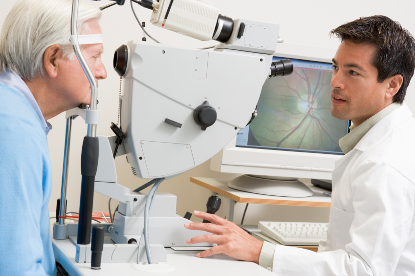
To see the internal structures of your eyes, the doctor will next ask you to rest your chin on a chinrest and press your forehead against a strap, while he aims an instrument at you called a slit lamp. The slit lamp is both a high-powered microscope and a light source that is focused to form a flat sheet. Because the front parts of the eye are fairly transparent, the sheet of light can show a cross section of the front structures of the eye, the way a sunbeam shining across a room can show the dust in the air. Depending on the width of the light beam and the lens, the slit lamp can give a magnified, three-dimensional view of the cornea, the iris, or the lens, or it can show a cross section from front to back of the eye, through the cornea, aqueous humor, lens, and vitreous. With an additional lens (either a handheld lens or one that fits directly against the cornea), the doctor can see all the way to the retina, blood vessels, and optic nerve at the back of the eye.
Another instrument used to view the interior of the eye and the retina is the ophthalmoscope. The most familiar type of ophthalmoscope is the handheld direct ophthalmoscope, which looks like a flashlight. Doctors use it to see the central retina. They may also use an indirect ophthalmoscope, which is a head-mounted instrument like a coal-miner’s lamp that shines into the eye and condenses the out-coming light into a three-dimensional image of the retina. Looking through the lens of the instrument and a handheld lens held in front of the patient’s eye, the doctor sees a wide, panoramic view of the retina.
To obtain the best view with the indirect ophthalmoscope — and sometimes with the slit-lamp — the doctor will first dilate your pupils with eyedrops, a procedure that may be unpleasant but not painful. Because your pupils may still be dilated for some time, it’s a good idea to bring a pair of sunglasses and make arrangements for transportation after the exam.
To the person having the eye exam, the standard tests may just seem like a barrage of bright lights. But to the eye doctor, they provide invaluable information. Here are the main conditions that might be spotted during the course of the exam and some of the ways they are treated.
Glaucoma
The vitreous and aqueous humor (the gelatinous and liquid substances that fill the eye) create intraocular pressure, a positive pressure inside the eye. Like the air inside a balloon, pressure in the eye maintains a smoothly rounded surface at the front of the cornea. Without this pressure, the eye surface would be rumpled — not a good optical lens.
If pressure in the eye becomes too high, however (due to poor drainage of the aqueous humor), it presses against the optic nerve, slowly restricting blood supply and killing the nerve cells. This is the general definition of glaucoma, although it has several distinct types. Glaucoma initially creates blind spots in the peripheral vision, but if it progresses untreated, it can cause blindness. Because peripheral vision is affected first, the field-of-vision test is an important way to screen for early glaucoma. Testing eye pressure is another.
Intraocular pressure is measured by briefly touching the eye with a tonometer. An applanation tonometer is attached to the slit lamp. You will see a mysterious-looking blue light that seems to come closer and closer to the eye, until it actually does touch the front of the eye. It cannot be felt, however, because the doctor applies a drop of local anesthetic before the test. The Tonopen is a smaller, handheld version of the applanation tonometer. There is also an air puff tonometer, which shoots a jet of air at the eye and often makes people jump.
It used to be said that normal intraocular pressure is exactly 21 millimeters of mercury, and that any pressure above that indicated glaucoma. In retrospect, that was as rigid as saying that normal body temperature is precisely 98.6°F. We now know that normal eye pressure varies from person to person. Many people with eye pressure in the low twenties do not have glaucoma, while some people with glaucoma have eye pressure in the high or even mid-teens.
The diagnosis of glaucoma is not made by eye pressure alone, but by characteristic changes in the appearance of the optic nerve and in the visual field. If eye pressure is normal but the visual field is reduced, the doctor may suspect glaucoma. Some people are marked “glaucoma suspects” for a while, meaning that it’s not absolutely clear that they have the disease. If a diagnosis cannot be made definitively, it’s reasonable to wait to begin treatment; if it turns out that the person does have glaucoma, it is still early enough that it has done no detectable damage. As soon as damage is noticed, though, treatment must be started immediately.
Treatment usually begins with medicated eyedrops, although some drugs can be taken orally. There are some kinds of glaucoma that require surgical or laser procedures. A laser procedure also may become necessary if eyedrops alone are not working well enough.
Cataract
When we are young, the lens of the eye is almost perfectly transparent, but during the normal course of aging, it gradually changes color and becomes cloudy. A clouded or opaque lens is called a cataract. There is no definite dividing line between normal aging changes and early cataract. Most people develop some cataract as they get older; people with diabetes are more likely than others to have cataract.
If a cataract gets bad enough to interfere with a person’s lifestyle and abilities, it can be removed with surgery and replaced with an intraocular lens, or implant. It used to be said that a cataract should be removed when it was “ripe,” but the decision to remove a cataract is really based on how much disability it causes a person. If it does not significantly impair your vision, it’s OK to wait. (It is possible to wait too long to have a cataract removed, but that is after the person can no longer see through it.) Cataract surgery is so quick and successful these days that there is no reason at all to dread or avoid it.
Retinal detachment
An eye problem that cannot wait to be treated is a retinal detachment. Most of eye’s interior is filled with vitreous, a gelatinous substance with fibers and cells that is firmly attached to the retina. Over time, the vitreous gradually becomes more liquid and less gelatinous. As the gelatin liquefies, the thin membrane at the back of the vitreous (the posterior vitreous membrane) can peel away from the retina and flap into the vitreous. This is called a posterior vitreous detachment, or PVD. It can occur without the person noticing it; alternatively, it can create dramatic flashes of light inside the eye, or brown or black floaters, which are actually the shadows cast on the retina either by the posterior vitreous membrane itself or by some leakage of blood into the vitreous.
While a PVD itself is not harmful, if part of the membrane is caught or has scarred to a spot on the retina, it can tear a hole in the retina as it peels away. If liquid vitreous then leaks through the hole and under the retina, the retina itself starts to separate from the outside of the eye. This is called a retinal detachment. Gradually, a person will lose sight in that eye, as if a curtain were slowly being drawn over it.
Most posterior vitreous detachments do not lead to retinal detachment, and many small retinal holes can be treated easily with a laser or even left alone. Because of the seriousness of a retinal detachment, however, anyone who notices new flashes and floaters needs to have an exam of the retina promptly. Fixing a retinal detachment requires complicated surgery; it is much better to treat it sooner rather than later.
Macular degeneration
The part of the retina responsible for central vision and fine visual acuity is the macula. Aging changes in the macula, called age-related macular degeneration, are one of the most common eye problems and the one that people seem to dread the most. Macular degeneration is caused by the breakdown of an insulating, outer layer of the retina. In the common, “dry” stage, the light-converting cells in the macula slowly deteriorate. In the less common but more serious “wet” form, harmful elements in the blood are able to reach the retina, damaging and scarring it.
Macular degeneration never causes total blindness, because it does not affect peripheral vision, but it makes images in the central vision appear extremely warped or blurry, and activities like driving a car or reading become difficult or impossible.
Good nutrition, exercise, adequate zinc intake, and not smoking are thought to prevent or slow the progression of macular degeneration, and many people with early macular degeneration continue to see well enough to lead a normal life. There are also some laser treatments, as well as two injectable medicines — ranibizumab (brand name Lucentis) and aflibercept (Eylea) — for the most severe kind of macular degeneration.
If vision loss reaches the level at which a person can no longer read with ordinary glasses, optical magnification systems and closed-circuit TV systems (although they are bulky and rather expensive) provide even greater magnification. When print is magnified, you only see a small area of what you are reading at any one time. This can take a little getting used to, but it is far better than being unable to read.
Diabetic retinopathy
The main reason people with diabetes are encouraged to have regular eye exams is to look for changes in the blood vessels of the retina that indicate diabetic retinopathy. Diabetic retinopathy develops as prolonged exposure to high blood glucose weakens the walls of the blood vessels in the eyes. The longer a person has had diabetes and the greater his exposure to high blood glucose, the greater his risk of having this condition.
The earliest signs of diabetic retinopathy are little red or white spots on the retina that can only be seen by an eye doctor. These spots are microaneurysms, tiny pouches of blood that have bulged through the damaged blood vessel walls and can leak blood, fat, and fluid into the retinal tissues. These early changes in the blood vessels are called background, or nonproliferative, retinopathy. Leaking in the retina from background retinopathy can cause some blurring of vision, but it does not usually require immediate treatment.
If diabetic retinopathy causes damage near the macula, however, fluid leaking into the macula makes it swell like a mosquito bite. This swelling, called macular edema, is the most common cause of visual impairment in diabetic retinopathy. (Reduced blood supply to the macula is a less common occurrence. As yet, it is not treatable.)
The earliest changes of retinopathy can even be temporary: here today and gone in six months. If damage continues, though, the risk increases that new, abnormal blood vessels will start to sprout in retina and poke through into the vitreous. This important change is called going from background retinopathy to proliferative retinopathy.
The new blood vessels apparently proliferate in an attempt to increase blood and oxygen supply to the damaged retina. They are so fragile, though, that they rupture at a cough, a sneeze, or even during sleep. Blood pours into the retina, blocking vision suddenly. When the bleeding stops, scar tissue forms, tugging at the retina and adding the potential for a retinal detachment.
Untreated proliferative retinopathy usually leads to blindness. Not long ago, diabetic retinopathy was just about the most discouraging condition that ophthalmologists had to deal with. Fortunately, in recent years, a number of large national studies have proven that laser treatment of the retina can help control proliferative diabetic retinopathy and significantly prolong useful vision.
Ironically, laser treatment works by producing scarring on the affected retina. The treated part of the retina will no longer see, but the laser treatment halts the growth of new blood vessels and preserves vision in the most important part of the retina, the macula. Laser treatment for diabetic retinopathy is a tremendous tool that has expanded the ability to treat diabetic eye problems.
A surgical procedure called a vitrectomy, in which blood, scar tissue, and vitreous are removed from the eye and replaced with a saline solution, is also a relatively new, successful treatment for retinopathy that affects the vitreous. The medicines ranibizumab and aflibercept are also approved to treat diabetic retinopathy in people with diabetic macular edema.
Looking out for your eyes
Currently, the American Diabetes Association recommends that adults and children 10 and older with Type 1 diabetes have an initial dilated and comprehensive eye examination within five years after they are diagnosed with diabetes. People with Type 2 diabetes are advised to have a dilated and comprehensive exam shortly after the diagnosis of diabetes. After the initial exam, everyone with diabetes is encouraged to have a yearly exam.
In a study published in the March 2001 issue of Diabetes Care, however, researchers studying retinal photographs taken during the Diabetes Control and Complications Trial discovered that in people with Type 1 diabetes, the progression of diabetic retinopathy begins even earlier than had previously been thought. Waiting up to five years to have an initial eye exam is too long, they suggested, because it may already be too late for the best treatment. They concluded that all individuals newly diagnosed with Type 1 diabetes should have an eye exam upon diagnosis.
With recent improvements in treatments, the chances of avoiding vision loss from diabetes- and age-related eye conditions are greater than ever, but vigilance is key. If you have never had a dilated eye exam or haven’t had one within the past year, ask your doctor for a referral to an eye-care professional who is trained to diagnose diabetic retinopathy. Take this important step now, and follow up with yearly visits.